sd lm fkfds mdskfs d sdlmfkfds mdskfsd sd lmfkfds mdskfsd sdlm fkfds mds kfsd sdl mfkfds mdsk fsd s dlmfkf ds mdsk fsd sdl mfkfds md skfs d sdlmfk fds mds kfsd sdlmfkfds mdsk fsd sdlm fkfds mdskf sd sd lmf kfds mdskf sd sd lmfkf ds mdskfsd s dlmfk fds m dskfsd sdlmfkf ds
							import imhr
							roi = imhr.eyetracking.ROI(isMultiprocessing=False, isDebug=True,
								# roi detection method and contour shape
								detection='manual', shape='straight',
								# path to read and export data
								image_path='./images', output_path='./output', metadata_source='metadata.xlsx',
								# stimuli size and screen size
								screensize=[1920,1080], recenter=[(1920*.50),(1080*.50)])
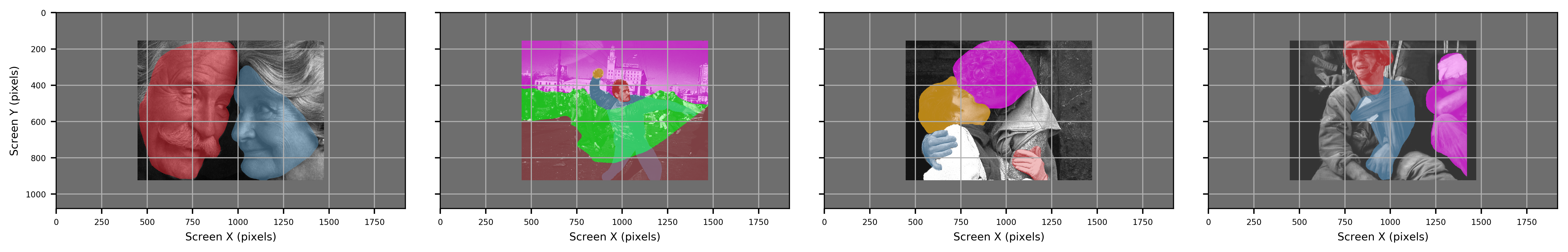
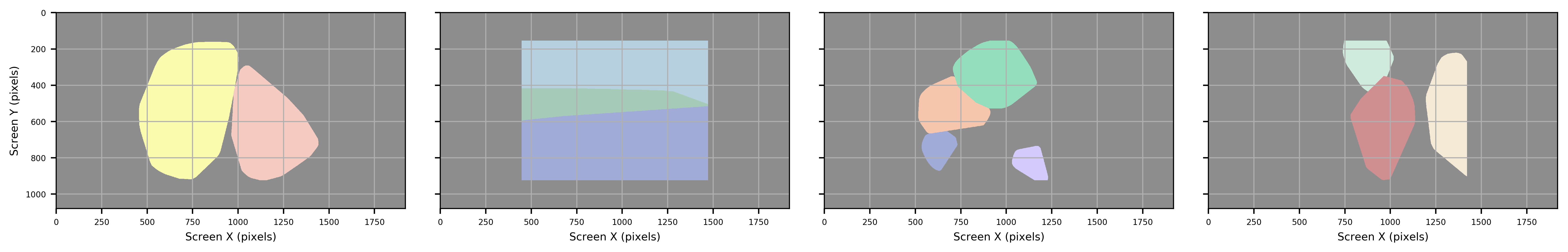
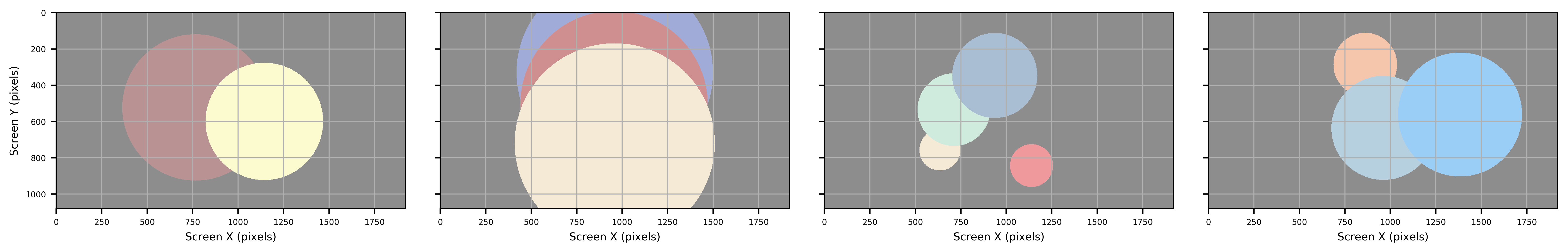
imhr.eyetracking.ROI() can identify Region/Area of Interest using machine learning and image coding techniques.
Defining Region of Interest
With the imhr library, there are two methods of creating regions of interest using manually highlighted images or haar cascades.
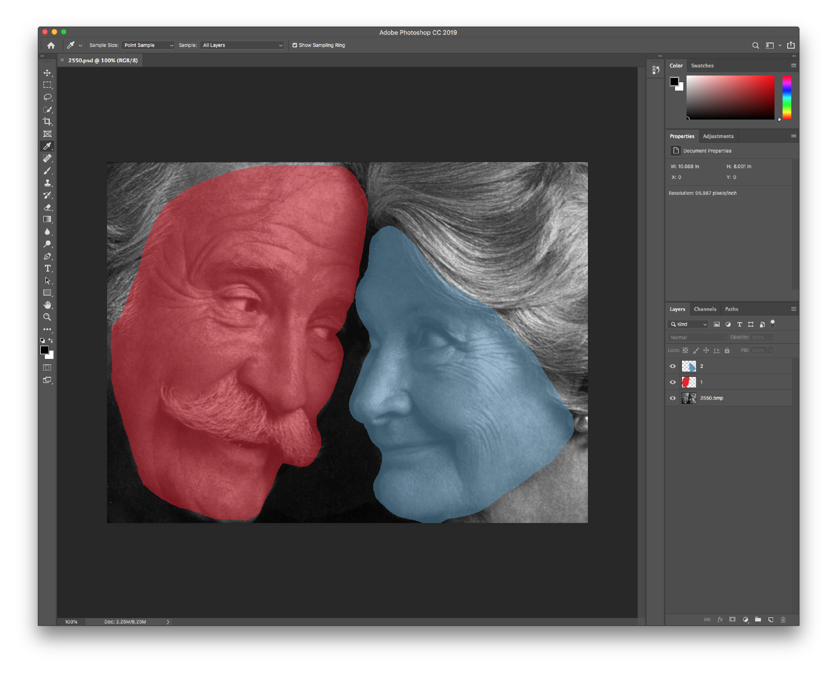
Manual highlighting
The first method is manually highlighting each region of interest by using a image media file that is able to store multiple layers of content. The two file types supported here are Adobe Photoshop Document (psd) and Digital Imaging and Communications in Medicine (dcm) filetypes. Each ROI can be stored as a seperate layer, which can then processed for exporting.
sd lm fkfds mdskfs d sdlmfkfds mdskfsd sd lmfkfds mdskfsd sdlm fkfds mds kfsd sdl mfkfds mdsk fsd s dlmfkf ds mdsk fsd sdl mfkfds md skfs d sdlmfk fds mds kfsd sdlmfkfds mdsk fsd sdlm fkfds mdskf sd sd lmf kfds mdskf sd sd lmfkf ds mdskfsd s dlmfk fds m dskfsd sdlmfkf ds
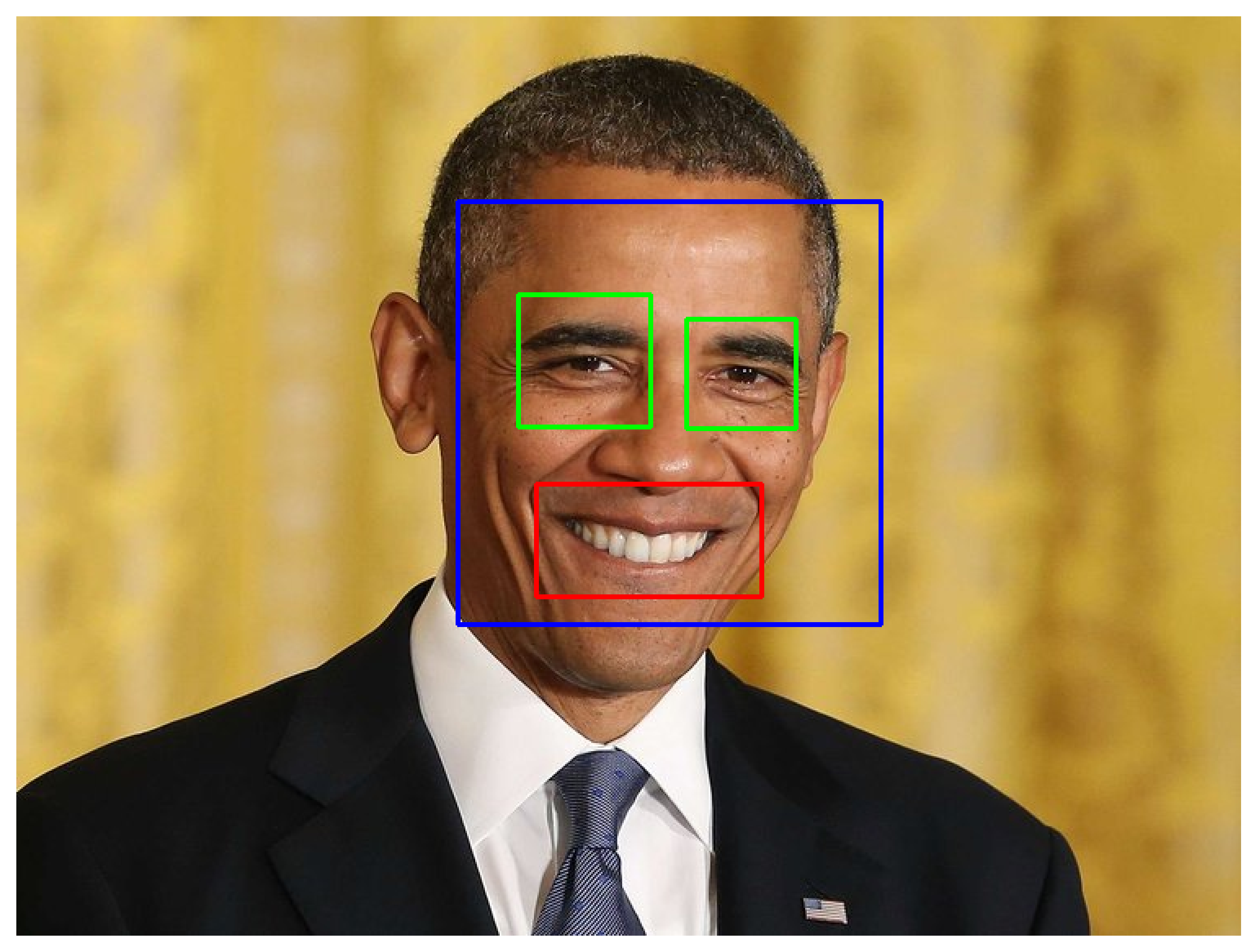
Haar Cascades
sd lm fkfds mdskfs d sdlmfkfds mdskfsd sd lmfkfds mdskfsd sdlm fkfds mds kfsd sdl mfkfds mdsk fsd s dlmfkf ds mdsk fsd sdl mfkfds md skfs d sdlmfk fds mds kfsd sdlmfkfds mdsk fsd sdlm fkfds mdskf sd sd lmf kfds mdskf sd sd lmfkf ds mdskfsd s dlmfk fds m dskfsd sdlmfkf ds
sd lm fkfds mdskfs d sdlmfkfds mdskfsd sd lmfkfds mdskfsd sdlm fkfds mds kfsd sdl mfkfds mdsk fsd s dlmfkf ds mdsk fsd sdl mfkfds md skfs d sdlmfk fds mds kfsd sdlmfkfds mdsk fsd sdlm fkfds mdskf sd sd lmf kfds mdskf sd sd lmfkf ds mdskfsd s dlmfk fds m dskfsd sdlmfkf ds
Bounds
sd lm fkfds mdskfs d sdlmfkfds mdskfsd sd lmfkfds mdskfsd sdlm fkfds mds kfsd sdl mfkfds mdsk fsd s dlmfkf ds mdsk fsd sdl mfkfds md skfs d sdlmfk fds mds kfsd sdlmfkfds mdsk fsd sdlm fkfds mdskf sd sd lmf kfds mdskf sd sd lmfkf ds mdskfsd s dlmfk fds m dskfsd sdlmfkf ds
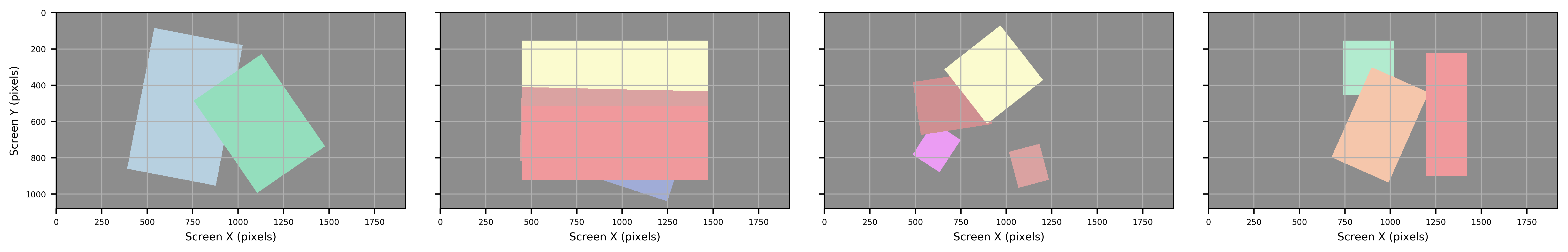
sd lm fkfds mdskfs d sdlmfkfds mdskfsd sd lmfkfds mdskfsd sdlm fkfds mds kfsd sdl mfkfds mdsk fsd s dlmfkf ds mdsk fsd sdl mfkfds md skfs d sdlmfk fds mds kfsd sdlmfkfds mdsk fsd sdlm fkfds mdskf sd sd lmf kfds mdskf sd sd lmfkf ds mdskfsd s dlmfk fds m dskfsd sdlmfkf ds
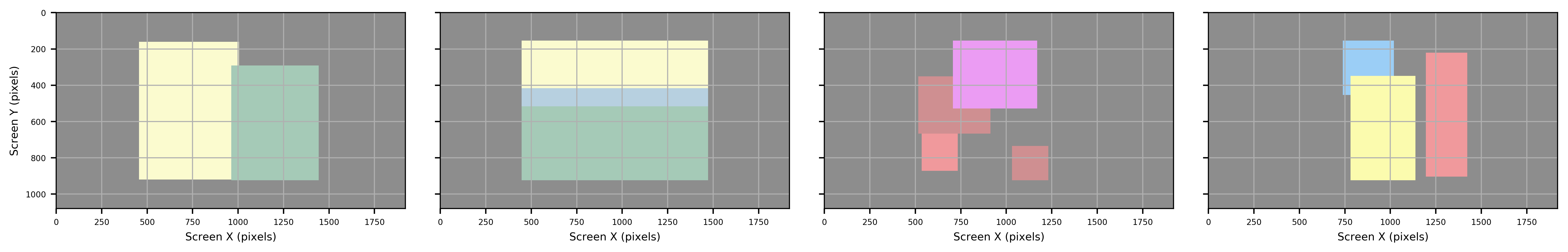
Output
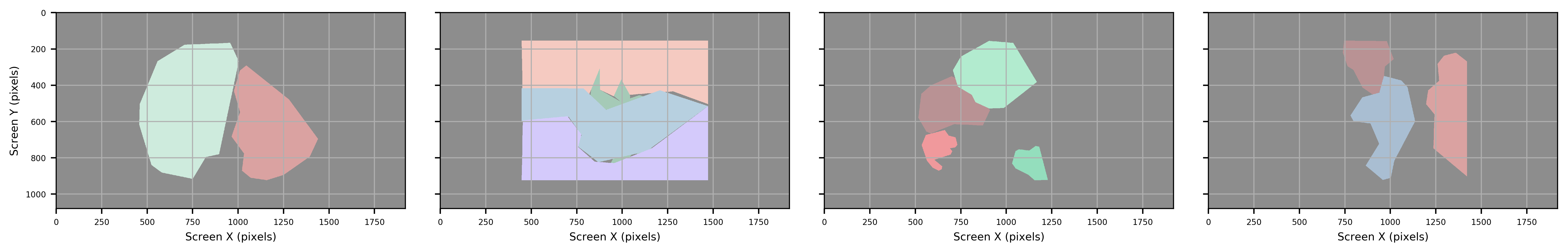
Two types of data are exported, contour shape and boundaries. Boundaries are saved in SR Research DataViewer (ias), and Microsoft Office Open XML Workbook (xlsx) formats, while contour shapes are saved in Hierarchical Data Format (HDF5; h5). Exported data can read either by SR Research DataViewer (ias) or any other data processing tool (xlsx, h5).
Both xlsx and HDF5 provide all metadata associated with each image, while ias files matches the format required by DataViewer. Of the three filetypes, HDF5 files are the most detailed providing both metadata for each ROI as well as exact pixel coordinates associated with each ROI.